Date and Location
Date: April 8, 2019 at 5 p.m. in the La Plata Multipurpose Room
Speaker: Hannah Horng
Background
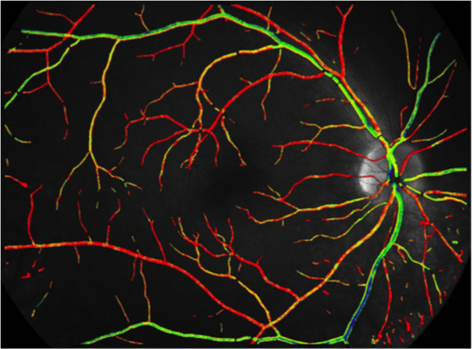
Retinal oximetry is a non-invasive technique that uses imaging methods to calculate the oxygen saturation of retinal blood vessels in the eye. Knowledge of these oxygen levels can allow the diagnosis of several different retinal diseases, like macular degeneration and tears in the retina. Imaging, however, requires the knowledge of the intricate network of veins and arteries that run through it, or its “vasculature.” In order to get an accurate representation of retinal vasculature, researchers in this experiment currently use a Photonic professional 3D printer to create models that can be used and analyzed.
Observations
The replica of the vasculature in the retina is called a “phantom,” as it represents a model of what is actually present in the eye. Via a 3D printing process, a laser is used to “write a solid” into a curable photoresist resin. The resolution is extremely high and is able to capture all the twists and turns of the branching vasculature, creating an empty “phantom” of the network. Once made and set, sample blood is injected into the phantom and the oxygen saturation levels are measured using the model.
One challenge the speaker emphasized that she and her research group faced was that during the creation of the phantom, some leftover resin would remain in the mold, as it remained undissolved with the appropriate solvent. This caused some of the hollow spaces within the vasculature to be filled with resin, which then caused issues later when blood was to be injected into the phantom. However, this challenge was overcome when they found that adding clear titanium dioxide would cause little to no excess resin to be leftover, allowing for better oxygen level analysis.
Again, it was evident that the speaker was fully interested in her subject of research, and went on to state that the use of 3D printing in the world of scientific research is something of great interest to her, and she believes it is the future of research itself.

Reflection and Questions
Overall, this was another great seminar. The speaker was invested in her topic and helped her audience learn more about her own field of research. While the presentation itself was engaging and interesting, the use of several unfamiliar terms and concepts (ex: multispectral imaging, planar retina model) made it a little difficult to keep and follow along during the seminar. Because of this, I would assume that her seminar was aimed more towards an audience already familiar with retinal oximetry and ophthalmology in general.
Questions:
- Do you think that 3D printing of phantoms can be used to model vasculature of other more complex systems within the human body?
- How exactly are oxygen saturation levels in the blood measured after injection into the phantom?
- How much time goes into the making of each phantom model? Is the process very tedious?


